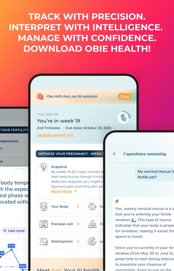
There are many parts of the uterus, placenta, amniotic fluid, and the fetus examined during the anatomic ultrasound examination around 20 weeksa of the pregnancy.
a. Fetal cardiac activity, fetal number, and presentation
b. A qualitative or semiquantitative estimate of amniotic fluid volume should be reported.
c. Placenta
The placental location, appearance, and relationship to the internal cervical os should be recorded. The umbilical cord should be imaged, and the number of vessels in the cord should be evaluated when possible.
d. Gestational (menstrual) age assessment.
First-trimester crown-rump measure- ment is the most accurate means for sonographic dating of pregnancy. Beyond this period, a variety of sono- graphic parameters such as biparietal diameter, abdominal circumference, and femoral diaphysis length can be used to estimate gestational (menstru- al) age. The variability of gestational (menstrual) age estimations, however, increases with advancing pregnancy. Significant discrepancies between gestational (menstrual) age and fetal measurements may suggest the possi- bility of a fetal growth abnormality, intrauterine growth restriction, or macrosomia.
or comparison.
i. Biparietal diameter (BPD)
Measured at the level of the thalami and cavum septi pellucidi. The cerebellar hemispheres should not be visible in this scanning plane. The measurement is taken from the outer edge of the proximal skull to the inner edge of the distal skull.
The head shape may be flattened (dolichocephaly) or rounded (brachycephaly) as a normal variant. Under these circum- stances, certain variants of normal fetal head development may make measurement of the head circumference more reliable than biparietal diameter for estimating gestational (menstrual) age.
ii. Head circumference (HC)
HC is measured at the same level as the biparietal diameter, around the outer perimeter of the calvarium. This measurement is not affected by head shape.
iii. Femoral diaphysis length (FL)
FL can be reliably used after 14 weeks’ gestational (menstrual) age. The long axis of the femoral shaft is most accurately measured with the beam of insonation being perpen- dicular to the shaft, excluding the distal femoral epiphysis.
iv. Abdominal circumference (AC)
AC should be determined at the skin line on a true transverse view at the level of the junction of the umbilical vein, portal sinus, and fetal stomach when visible. AC measurements are used with other biometric parameters to estimate fetal weight and may allow detection of intrauterine growth restriction or macrosomia.
e. Fetal weight estimation.
Fetal weight can be estimated by obtaining measurements such as the biparietal diameter, head circum- ference, abdominal circumference or average abdominal diameter, and femoral diaphysis length. Results from various prediction models can be compared to fetal weight per- centiles from published nomograms.
f.Maternal anatomy.
Evaluation of the uterus, adnexal structures, and cervix should be performed when appropriate. When the cervix cannot be visualized, a transperineal or transvaginal scan may be considered when evaluation of the cervix is needed.
g. Fetal anatomic survey.
Fetal anatomy may be adequately assessed by ultrasound after approximately 18 weeks’ gestational (menstrual) age. It may be possible to document normal structures before this time, although some structures can be difficult to visualize because of fetal size, position, movement, abdominal scars, and increased maternal abdominal wall thickness.
A second- or third-trimester scan may pose technical limitations for an anatomic evaluation because of imaging artifacts from acoustic shad- owing. When this occurs, the report of the sonographic examination should document the nature of this technical limitation. A follow-up examination may be helpful.
The following areas of assessment represent the minimal elements of a standard examination of fetal anatomy. A more detailed fetal anatomic examination may be necessary if an abnormality or suspected abnormality is found on the standard examination.
i. Head, face, and neck Cerebellum
Choroid plexus Cisterna magna Lateral cerebral ventricles Midline falx Cavum septi pellucidi Upper lip. A measurement of the nuchal fold may be helpful during a specific age interval to suggest an increased risk of aneuploidy.
ii. Chest
The basic cardiac examination includes a 4-chamber view of the fetal heart. If technically feasible, views of the outflow tracts should be attempt- ed as part of the cardiac screening examination.
iii. Abdomen: Stomach (presence, size, and situs); Kidneys; Bladder; Umbilical cord insertion site into the fetal abdomen; Umbilical cord vessel number
iv. Spine: Cervical, thoracic, lumbar, and sacral spine
v. Extremities: Legs and arms: presence or absence
vi. Sex: Medically indicated in low-risk pregnancies only for evaluation of multiple gestations.