First Trimester Ultrasound Scanning Before 8-10 Weeks
Ultrasound
Obie Editorial Team

The ultrasound before 8-10 weeks
In the first trimester, even at the first prenatal visit, ultrasound scanning during pregnancy is often useful to identify many pregnancy and fetal abnormalities and it also provides an accurate dating of a pregnancy. First trimester scanning can be performed using either an abdominal approach or a vaginal approach. The sonogram done before 8-10 weeks is different from the first trimester screen which is a sonogram that measures the fetus' nuchal fold plus a blood test.
Abdominal ultrasound scanning is usually performed with a full maternal bladder, provides a wider field of view, and the greatest depth of view. Vaginal scanning is best performed with the bladder empty, gives a much greater resolution with greater crispness of fine detail. In circumstances where both approaches are readily available, the greater detail provided by transvaginal scans usually outweighs other considerations, and is preferred.
Scanning is usually done in the normal examination position (dorsal lithotomy) with her feet secure in stirrups and her perineum even with the end of the examination table.
The following findings can be made during early first trimester ultrasound sonography:
- Development of a pregnancy
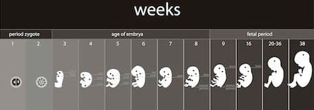
- Determination of gestational age
- Miscarriage
- Ectopic pregnancy
- One or more fetuses
- Ovarian assessment (eg follicle)
- Nuchal thickness
What can you see before 10 weeks of pregnancy by ultrasound?
There are several benefits to ultrasound evaluation before 8-10 weeks:
- Detection of abnormal pregnancies that are destined to miscarry.
- Enabling scheduled intervention, if desired by the patient.
- Enabling collection of pregnancy tissue for chromosomal analysis, if desired by the patient.
- Reassurance to the patients with normal ultrasound scans.
This is what can be seen and often measured before 10 weeks of the pregnancy:
- Intrauterine versus ectopic pregnancy
- Gestational Sac
- Yolk Sac
- Fetal heart beat
- Fetal Pole
- Crown Rump Length
- Number of fetuses
In the presence of uterine bleeding, visualization of a gestational sac, a yolk sac, a fetal pole and fetal heart beat changes the risk of a threatened abortion leading to miscarriage from 50/50 to about 5%.
Observation of subchorionic bleeding (blood outside the sac) is noted in about 20% of patients with threatened abortion. This is a potentially worrisome sign, and reduces the pregnancy continuation rate to about 2/3.


Read More